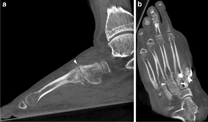
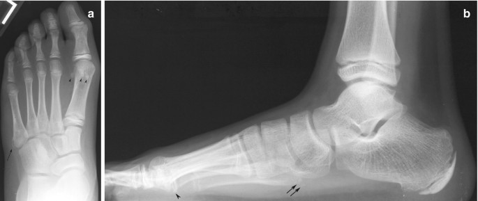
Foot X-ray of a 10 year-old male patient (white arrow indicates
5 (129) · $ 22.50 · In stock
Imaging of osteoarthritis from the ankle through the midfoot
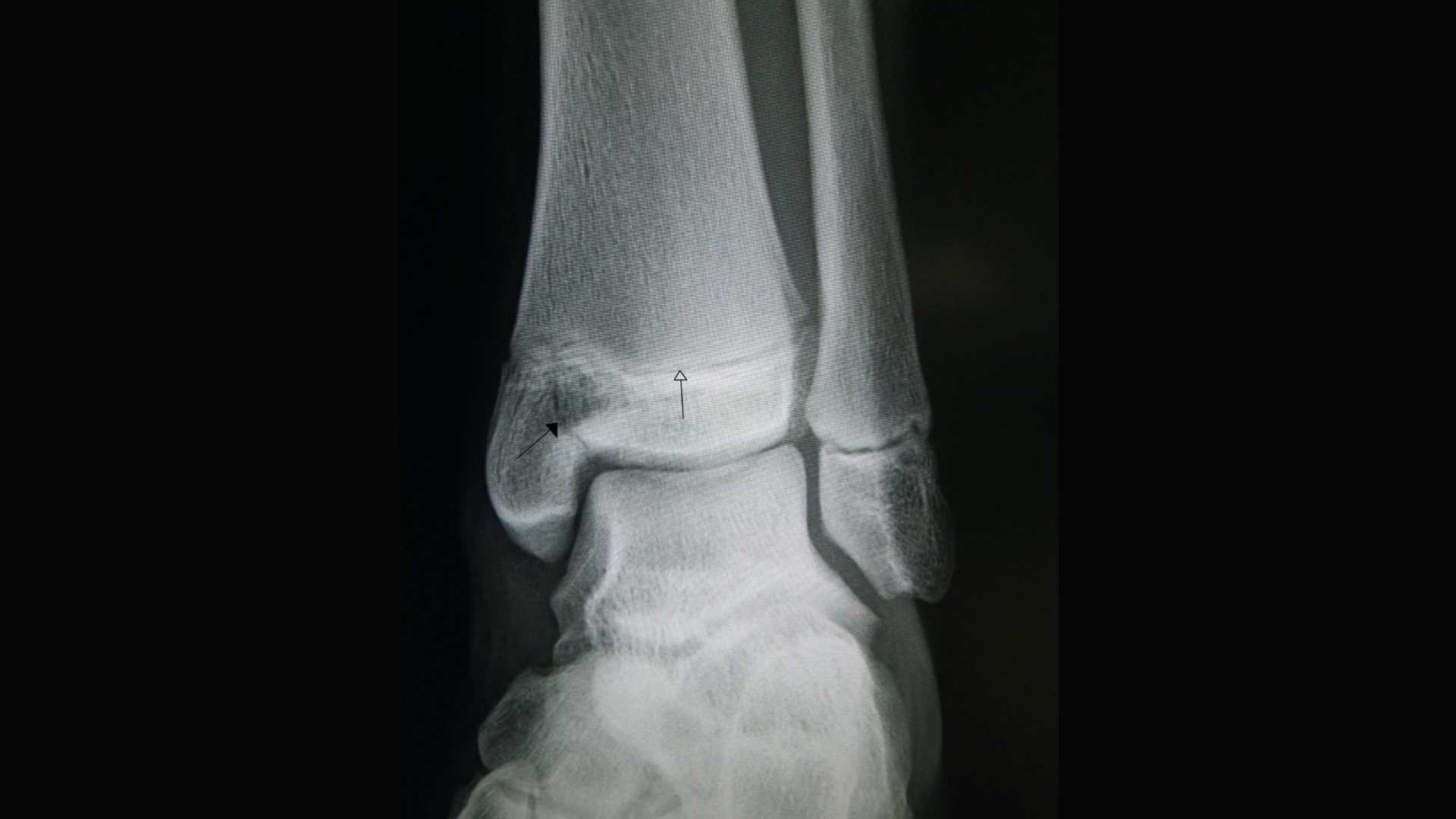
Musculoskeletal (MSK) X-ray Interpretation - OSCE Guide
JCM, Free Full-Text
![Soyuz Drop Test Platform [24] Figure 3-9: Seat Testing at Various](https://www.researchgate.net/profile/Jamiu-Omirinde/publication/269802154/figure/fig2/AS:295007710924803@1447346727496/B-Amelioration-of-radiation-induced-histological-changes-in-testes-of-Wistar-rats-at-day_Q320.jpg)
Soyuz Drop Test Platform [24] Figure 3-9: Seat Testing at Various
Radiology of the Pediatric Foot and Ankle

SciELO - Brasil - Unusual imaging characteristics of thoracic hydatid disease Unusual imaging characteristics of thoracic hydatid disease

Ozlem Bilir's research works Recep Tayyip Erdoğan Üniversitesi, Rize and other places

Normal ankle x-ray - 11-year-old, Radiology Case
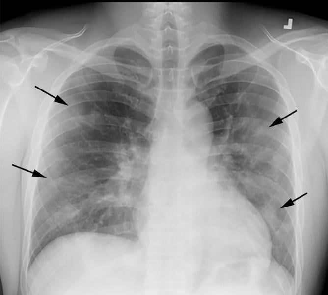
PHOTO GALLERY: How COVID-19 Appears on Medical Imaging
![Soyuz Drop Test Platform [24] Figure 3-9: Seat Testing at Various](https://www.researchgate.net/publication/259309727/figure/fig3/AS:669535099158531@1536641015064/8-Soyuz-Drop-Test-Platform-24-9-Seat-Testing-at-Various-Angles-24.jpg)
Soyuz Drop Test Platform [24] Figure 3-9: Seat Testing at Various
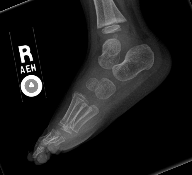
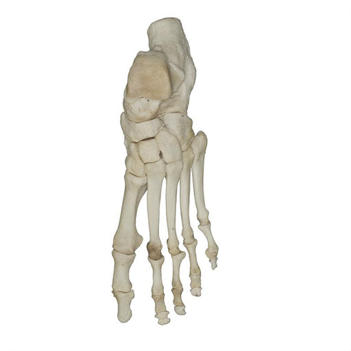
Normal Bones

Asim KALKAN, Professor (Full), Professor

Apls Pediatric Emergency Radiology 1
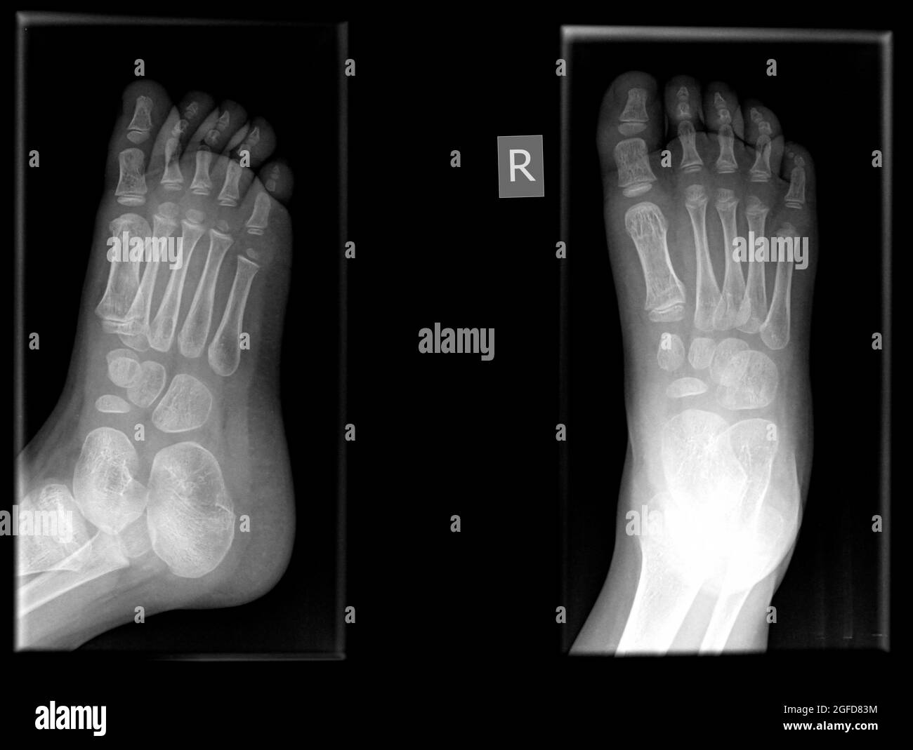
Radiograph foot hi-res stock photography and images - Alamy

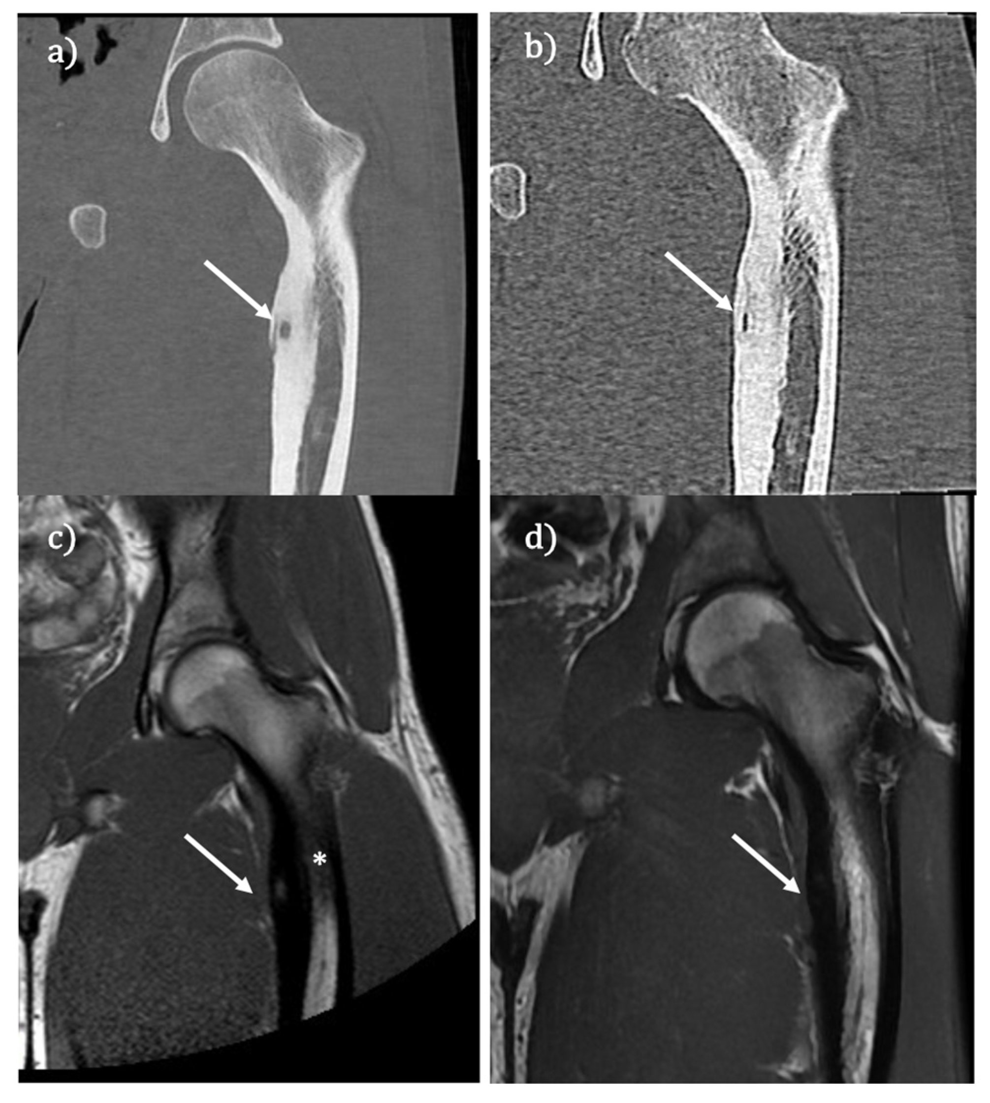
a) Lateral X-ray of the patient. (b) Red line (*) denotes a fracture