Cellular fluorescence intensity and cell size as a function of
4.7 (149) · $ 22.00 · In stock

Download scientific diagram | | Cellular fluorescence intensity and cell size as a function of enrichment substrate on a subset of basalt enrichments (sample 30R-1A). The circle size indicates the average cellular area (mm 2 ) as measured by epi-fluorescence microscopy. The standard deviation for the cellular area of each sample is roughly 46% based on the following number of cells counts for each treatment: Host Rock (n = 28); No addition (n = 41); CH 3 COONa (n = 38); CH 4 (n = 62); NaHCO 3 (n = 43); NaHCO 3 + NH 4 Cl (n = 39); NaHCO 3 + NaNO 3 (n = 36); NH 4 Cl (n = 35); NaNO 3 (n = 38). Symbols ++ and + indicate analysis of variance P-values of <0.01 and <0.05, respectively, versus Host rock or No addition; and * * indicate P < 0.01 versus CH 3 COONa, CH 4 or NaHCO 3 . from publication: Nitrogen Stimulates the Growth of Subsurface Basalt-associated Microorganisms at the Western Flank of the Mid-Atlantic Ridge | Oceanic crust constitutes the largest aquifer system on Earth, and microbial activity in this environment has been inferred from various geochemical analyses. However, empirical documentation of microbial activity from subsurface basalts is still lacking, particularly in the | Nitrogen, Stimulation and Geomicrobiology | ResearchGate, the professional network for scientists.

Antibodies 101: Flow Cytometry
Pulse-number-dependent fluorescence intensity and cell viability of the

Cellular uptake showing comparison of intracellular fluorescence of doxorubicin: Incubation of MDAMB231 cells with different doxorubicin formulations (1 µM) for 2 h is shown by (a) Confocal microscopy images showing intracellular fluorescence

PDF) Nitrogen Stimulates the Growth of Subsurface Basalt
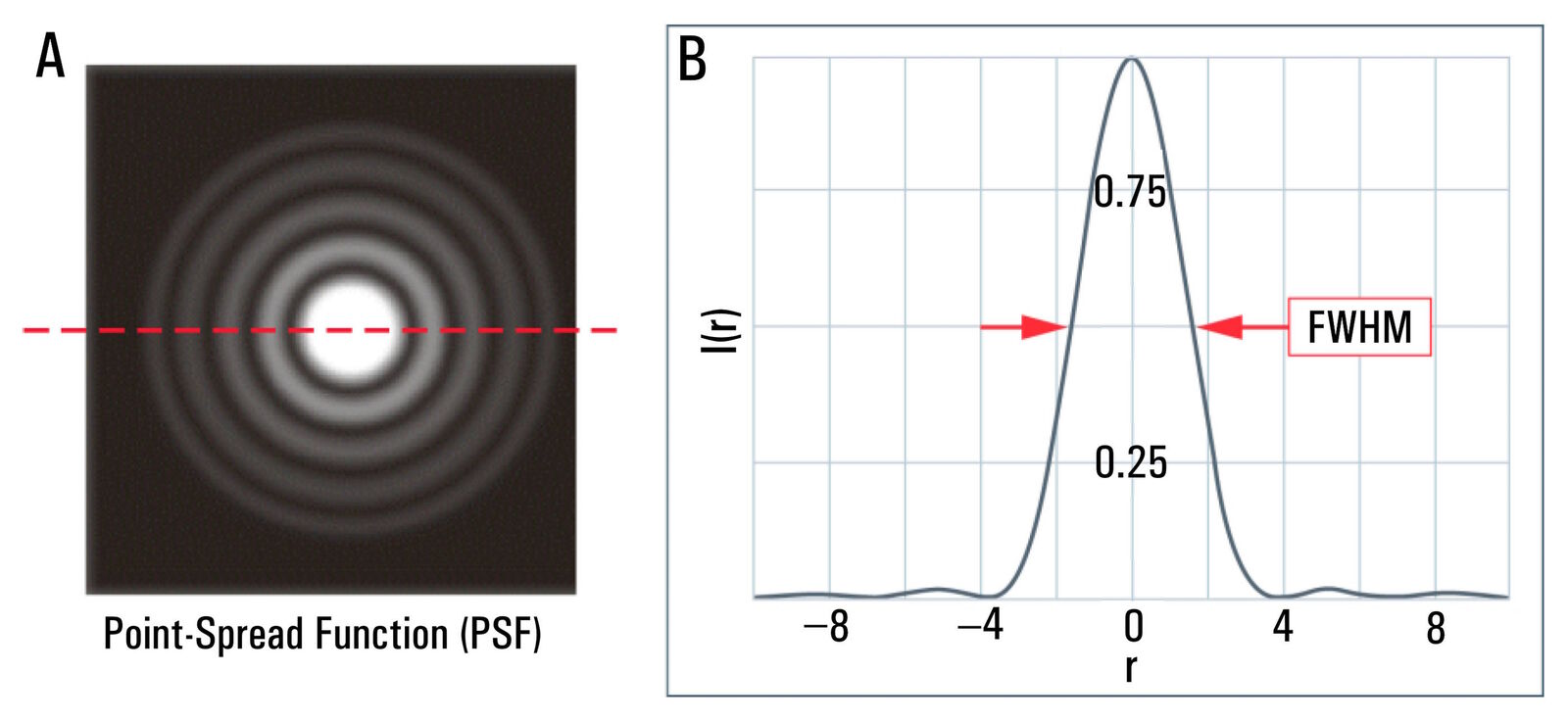
Microscope Resolution: Concepts, Factors and Calculation, Science Lab

Schematics of SAPS (successive alkalinity producing system

Variability of fluorescence intensity distribution measured by flow cytometry is influenced by cell size and cell cycle progression
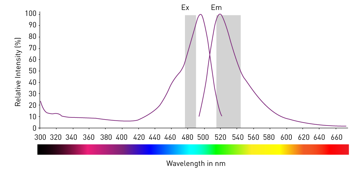
Fluorescence Intensity Measurements
Sensitivity of endogenous autofluorescence in HeLa cells to the application of external magnetic fields